Air Hunger: Why You Can't Get a Full Breath When Your Oxygen Is Perfect
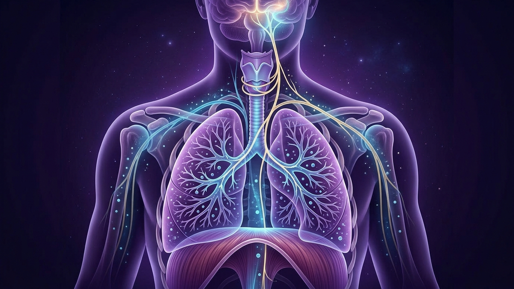
Few symptoms frighten a patient more than the feeling that they cannot get a full breath. They describe air that will not "catch," a chest that will not fill, a compulsion to sigh or yawn that never satisfies. They arrive with a normal chest X-ray, clear lungs, an oxygen saturation of 98 percent, and a physician who tells them nothing is wrong. But something is wrong. The conventional frame assumes breathlessness means the lungs are failing to oxygenate the blood. In the vast majority of these patients, oxygenation is flawless. What has failed is not the lung — it is the signaling loop between the airway, the vagus nerve, and the brain that decides whether a breath was enough. Air hunger is a disorder of respiratory perception, and its center is vagal.
Dyspnea Is Manufactured in the Brain, Not the Lung
The sensation we call breathlessness is not a direct readout of blood gases. It is a construction — assembled in the insula and limbic cortex from a comparison between two streams of information. The first is respiratory drive: the outgoing motor command the brainstem sends to the diaphragm and accessory muscles, along with a "corollary discharge" copy of that command sent up to sensory cortex. The second is the incoming afferent feedback reporting what the breath actually accomplished. When the outgoing command and the incoming feedback match, breathing feels effortless and drops below conscious awareness. When they diverge — when the brain has asked for a satisfying breath and the returning signal says that wasn't it — the mismatch is experienced as air hunger. This is the modern neurophysiological consensus: dyspnea is a mismatch signal, closely analogous to how pain is generated. And the single largest channel carrying that afferent feedback is the vagus nerve.
The Vagus Is the Lung's Sensory Reporter
Roughly 80 percent of vagal fibers are afferent — sensory fibers carrying information from the body up to the brainstem. The lungs are densely instrumented with vagal sensory endings, and three types matter here. Slowly adapting pulmonary stretch receptors sit in airway smooth muscle and fire as the lung inflates; their signals travel the vagus to the nucleus tractus solitarius and drive the Hering-Breuer reflex, the circuit that senses adequate inflation and terminates the inspiratory effort. This is the vagal system that tells the brain "the breath was full — you can let go." Rapidly adapting (irritant) receptors respond to mechanical distortion and inflammation and provoke the urge to cough, sigh, and take deeper breaths. Pulmonary C-fibers, unmyelinated vagal afferents near the alveoli, are exquisitely sensitive to inflammatory mediators and lung congestion and are potent generators of the raw sensation of air hunger.
Now consider what happens when vagal tone is low and vagal signaling is distorted — the state we see in dysautonomia, POTS, long COVID, ME/CFS, chronic anxiety, and post-viral syndromes generally. The stretch-receptor feedback that should say "full breath achieved" arrives weakened or mistimed. The Hering-Breuer "satisfaction" signal fails to close the loop. The brain keeps issuing the command for a fuller breath, the feedback keeps under-reporting, and the mismatch never resolves. The patient sighs, yawns, and gulps air precisely because the vagal signal that would tell them they succeeded is not landing. Air hunger, in this reading, is a vagal afferent reporting failure — a lung that is working normally but whose success is not being communicated upward.
The Chemoreflex and the CO2 Paradox
There is a second mechanism, and it is the one that turns air hunger into a self-perpetuating trap. Respiratory drive is governed less by oxygen than by carbon dioxide. Central and peripheral chemoreceptors — the latter carried, again, partly by vagal and glossopharyngeal afferents from the carotid and aortic bodies — set the urge to breathe based largely on arterial CO2 and pH. When a frightened, dysregulated person breathes a little too much, they blow off CO2. Arterial CO2 falls, blood pH rises into respiratory alkalosis, and here is the cruel paradox: low CO2 does not relieve air hunger, it can worsen it. Hypocapnia causes cerebral vasoconstriction (the dizziness and derealization), shifts the oxygen-hemoglobin dissociation curve so tissues actually release less oxygen despite full saturation, and drives the tingling, chest tightness, and limb paresthesias that patients read as further evidence of suffocation. So they breathe harder. Which lowers CO2 further. This is the engine of chronic hyperventilation syndrome, and most of these patients are not gasping visibly — they are subtly over-breathing all day, chronically hypocapnic, with a chemoreflex that has been recalibrated to defend an abnormally low CO2 set point. Every deep, effortful "rescue" breath deepens the disorder.
Why the Diaphragm Makes It Worse
Layered on top is a mechanical failure. Under chronic stress and low vagal tone, breathing migrates upward — out of the diaphragm and into the accessory muscles of the neck, shoulders, and upper chest. This apical, thoracic-dominant pattern is inefficient: it moves air into the upper lung, where stretch-receptor feedback is less satisfying, and it recruits muscles that fatigue and ache, feeding the sense of effort. When such a patient is told to "take a deep breath," they perform a large, forceful accessory-muscle inhalation that further drops CO2 and generates a large corollary discharge with disappointing stretch feedback — a bigger mismatch, more air hunger. This is why the intuitive fix makes it worse, and why the intervention must be counterintuitive: less air, slower, lower, through the nose.
How to Recognize a Vagal Breathing-Pattern Disorder
This phenotype has a recognizable clinical signature:
- The complaint is "I can't get a full breath" or "I can't get the breath to catch" — not true shortness of breath on exertion. It is frequently worse at rest and when attention turns inward.
- Frequent sighing, yawning, throat-clearing, and a feeling of needing to consciously breathe.
- Oxygen saturation is normal; lungs are clear; the workup is repeatedly negative.
- Accompanying tingling around the mouth or in the fingers, lightheadedness, palpitations, and a wave of anxiety — the fingerprint of hypocapnia.
- Symptoms fluctuate with emotional state and improve, tellingly, during absorbed distraction or sleep.
- Low resting heart rate variability and other markers of reduced vagal tone.
Red Flags: Rule Out the Dangerous Causes First
Air hunger with a normal workup is common and benign in mechanism — but the label must be earned, never assumed. Certain features demand urgent medical evaluation before anyone reframes the symptom as functional. Seek prompt care for: breathlessness of sudden onset, breathlessness with chest pain or pain radiating to the arm or jaw, breathlessness that is genuinely worse with exertion or when lying flat, a measured oxygen saturation that is actually low, coughing blood, unilateral leg swelling (a pulmonary embolism can present as air hunger with normal saturation and must be excluded), fever, or a known cardiac or pulmonary history. Anemia, thyroid disease, arrhythmia, heart failure, asthma, and PE all masquerade as this. A coach is not a diagnostician: the responsible move is always to ensure a clinician has cleared the cardiopulmonary causes first, and any changes to prescribed cardiac, respiratory, or anxiety medication belong with that clinician, never made alone. Only then does breathing retraining become the intervention.
Where Neuromodulation and Breath Retraining Fit
Air hunger is arguably the flagship condition for vagal breath retraining, because the intervention acts directly on the broken loop. The goal is not to breathe more. It is to restore vagal afferent feedback, raise CO2 tolerance, and rebuild the diaphragmatic pattern that generates satisfying stretch-receptor signals.
Slow, low, nasal breathing
Breathing at approximately six breaths per minute, through the nose, into the belly, with an exhale longer than the inhale, is the foundational intervention. The extended exhale directly increases vagal efferent output (respiratory sinus arrhythmia), while the slow, full diaphragmatic inflation restores the stretch-receptor feedback that closes the Hering-Breuer loop — the breath finally "registers." Nasal breathing matters: it adds resistance, slows the rate, and recruits nasal nitric oxide, which improves ventilation-perfusion matching.
CO2 tolerance work
Because the core lesion is a chemoreflex defending too-low a CO2, gentle tolerance work — comfortable reduced-volume breathing and short, unforced breath-holds after a normal exhale — retrains the brainstem to accept a higher, healthier CO2. Patients learn that the urge to breathe is a signal to be observed, not obeyed. This is interoceptive re-education as much as respiratory training.
Humming, gargling, and singing
Humming on the exhale is uniquely useful: it lengthens the exhale, vibrates the pharynx and larynx (richly vagally innervated), and dramatically increases nasal nitric oxide. Gargling and singing recruit the same vagal territory. These are, in effect, self-administered vagal maneuvers.
HRV biofeedback and the emerging tech
HRV biofeedback makes the vagal system visible, letting the patient find their personal resonance frequency and watch vagal tone climb in real time — steadying for someone convinced their body is failing. Beyond breath, transcutaneous auricular vagus nerve stimulation (taVNS) delivers mild electrical stimulation to the vagal-innervated outer ear, and early data suggest it can raise vagal tone and dampen the anxiety-hyperventilation loop. And low-intensity focused ultrasound aimed at the cervical vagus is an emerging, non-invasive way to modulate autonomic tone directly; it is being studied across dysautonomic and post-viral populations, and we expect the evidence base to mature over the coming years. These are adjuncts to breath retraining, not replacements for it.
Clinical takeaway: Air hunger with normal oxygen is not lung failure — it is a vagal-interoceptive signaling disorder driven by disrupted pulmonary stretch-receptor feedback, an over-sensitive chemoreflex defending chronically low CO2, and an accessory-muscle breathing pattern that makes every "deep breath" worse. Once the dangerous cardiopulmonary causes are excluded, the treatment is counterintuitive and reliable: slower, lower, nasal, exhale-led breathing to restore vagal tone and CO2 tolerance, supported by HRV biofeedback and emerging vagal neuromodulation. The patient who cannot get a full breath is not imagining it — their brain simply is not being told the breath already succeeded.
References
- Parshall MB, Schwartzstein RM, Adams L, et al. "An official American Thoracic Society statement: update on the mechanisms, assessment, and management of dyspnea." American Journal of Respiratory and Critical Care Medicine, 2012;185(4):435-452.
- Banzett RB, Lansing RW, Binks AP. "Air hunger: a primal sensation and a primary element of dyspnea." Comprehensive Physiology, 2021;11(2):1449-1483.
- Undem BJ, Kollarik M. "The role of vagal afferent nerves in chronic obstructive pulmonary disease and airway sensation." Proceedings of the American Thoracic Society, 2005;2(4):355-360.
- Boulding R, Stacey R, Niven R, Fowler SJ. "Dysfunctional breathing: a review of the literature and proposal for classification." European Respiratory Review, 2016;25(141):287-294.
- Gardner WN. "The pathophysiology of hyperventilation disorders." Chest, 1996;109(2):516-534.
- Lehrer PM, Gevirtz R. "Heart rate variability biofeedback: how and why does it work?" Frontiers in Psychology, 2014;5:756.
- Yuan H, Silberstein SD. "Vagus nerve and vagus nerve stimulation, a comprehensive review: part I." Headache, 2016;56(1):71-78.