Gastroparesis: The Clearest Case of Vagal Failure in Medicine
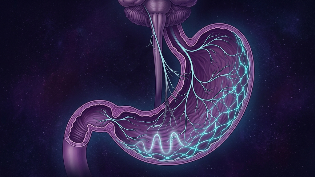
When a patient is told they have gastroparesis, the conversation almost always centers on the stomach — as if the muscle simply forgot how to squeeze. But the stomach is not an autonomous organ. It is a motor driven, moment to moment, by the vagus nerve. Gastroparesis is the single clearest, most nameable example of vagal failure in all of clinical medicine: a motor nerve losing its grip on the organ it is built to command. Once you see it that way, the early satiety, the bloating, the nausea, the vomiting of food eaten hours ago, and the erratic blood sugar stop looking like a mysterious stomach disease and start looking like exactly what they are — a nerve that has gone quiet.
The Vagus Is the Motor Nerve of the Stomach
Most people think of the vagus as a vague calming wire, the thing you "tone" with cold plunges. In the stomach it is something far more concrete: it is the wiring that makes the organ move. Gastric emptying is not one action but a choreographed sequence, and the vagus conducts every measure of it.
Three coordinated events have to happen for a meal to leave the stomach on schedule:
- Receptive relaxation and fundic accommodation. The moment you swallow, vagal fibers signal the upper stomach (the fundus) to relax and expand, making room for food without a pressure spike. This is a vagovagal reflex, mediated by inhibitory neurons that release nitric oxide. When it fails, even a small meal produces immediate, distressing fullness.
- The antral pump. The lower stomach (the antrum) grinds and propels food toward the outlet in rhythmic contractions. The vagus sets the strength and coordination of these contractions. Lose vagal drive and the antrum goes weak and disorganized — food is churned feebly instead of pushed forward.
- Pyloric coordination. The pylorus, the muscular valve at the stomach's exit, must open in time with each antral wave. Vagal input coordinates that timing. When coordination is lost, the pylorus can stay clamped — pylorospasm — while the antrum pushes against a closed door.
The vagus does not act on the muscle directly. It works through the stomach's own pacemaker network: the interstitial cells of Cajal (ICC). These specialized cells generate the slow electrical waves — roughly three per minute in the human stomach — that set the rhythm every contraction follows. The vagus is the conductor; the ICC are the metronome; the smooth muscle is the orchestra. In gastroparesis, both the conductor and the metronome are commonly damaged. Biopsy studies of gastroparetic stomachs repeatedly show depleted and disrupted ICC networks alongside signs of autonomic neuropathy. This is why the disorder is so stubborn — the failure is at the level of the control system, not the muscle fibers themselves.
How the Vagus Gets Injured
Gastroparesis is not one disease. It is a final common pathway that several very different injuries lead to, and naming which one a patient has matters for prognosis.
- Diabetic autonomic neuropathy — the classic cause. Chronic hyperglycemia damages small autonomic fibers, and the long, exposed vagus is among the most vulnerable nerves in the body. Diabetic gastroparesis is the textbook example, and it illustrates the cruel feedback loop described below.
- Post-viral vagal injury. This is the phenotype exploding in visibility right now. Viruses with neurotropic reach — Epstein-Barr, and now SARS-CoV-2 — can inflame or injure vagal fibers and the brainstem nuclei that feed them. Many long COVID patients with new, severe early satiety and nausea are describing post-viral gastroparesis, even when no one has named it. Post-EBV cases have been recognized for decades.
- Post-surgical vagal damage. The vagus runs directly through the surgical field of fundoplication, bariatric procedures, and esophageal operations. It can be stretched, cut, or entrapped. Gastroparesis that begins in the weeks after upper-abdominal surgery is often frank mechanical vagal injury.
- Idiopathic. A large share of cases have no identified cause — and many of these, on careful history, turn out to be post-viral events that were never connected to the illness that triggered them.
The diabetic and post-viral forms share a mechanism worth dwelling on because it explains the blood-sugar chaos patients describe. Gastric emptying is one of the largest determinants of the post-meal glucose curve. When emptying is delayed and erratic, glucose arrives late and unpredictably — rapid-acting insulin dosed for a normal meal peaks before the food does, causing a low, then a delayed high when the stomach finally empties. In diabetics this creates a vicious loop: high glucose itself further slows emptying and worsens the neuropathy, which worsens glycemic control. Breaking that loop anywhere — better vagal function, smaller meals, or insulin timing adjusted with a clinician — helps everywhere.
The Clinical Signature
Vagal-failure gastroparesis has a recognizable fingerprint, and coaches working alongside a medical team should learn to read it:
- Early satiety — feeling full after a few bites, the hallmark of lost fundic accommodation.
- Postprandial fullness and bloating that lingers for hours because the meal is not leaving.
- Nausea and vomiting of undigested or "old" food — food eaten hours earlier, sometimes overnight, still recognizable. This is close to pathognomonic and separates gastroparesis from ordinary indigestion.
- Reflux overlap. A stomach that will not empty pushes contents upward, so many patients carry a reflux diagnosis and a shelf of failed acid suppressants — the same incomplete-diagnosis pattern seen across vagal upper-GI disease.
- Erratic blood sugar and unintentional weight change, usually loss, sometimes gain from grazing on the few foods that stay down.
- Low resting heart rate variability — a systemic marker that the same vagal collapse is affecting the heart, often alongside POTS or broader dysautonomia.
To the Patient Told It Is "Vagus Nerve Damage"
If a clinician has told you that you have vagus nerve damage, that was not a dismissal — it was, for once, an accurate diagnosis. It means the nerve that runs your stomach is not signaling the way it should. That is worth grieving and then working with, because it also carries real hope.
Nerve injury is rarely all-or-nothing. Most gastroparesis is partial vagal dysfunction, not a severed cable. Partially injured autonomic fibers can recover function over months, especially in post-viral and post-inflammatory injuries, as inflammation resolves and surviving fibers take up slack. The ICC network, too, can regenerate under better metabolic conditions. Recovery is usually gradual and non-linear — good weeks and bad weeks — rather than a switch flipping. The goal of everything below is to protect the nerve you have, reduce the load it must carry, and give a partially injured system the best possible conditions to relearn its rhythm.
Where Neuromodulation and Ultrasound Fit
If the problem is a motor nerve that has gone quiet, the most mechanistically honest interventions are the ones that speak to that nerve directly.
Breath as the daily input
Slow diaphragmatic breathing at roughly six breaths per minute, with exhales longer than inhales, is the most reliable non-invasive way to raise vagal output — and the diaphragm's movement mechanically massages the stomach as a bonus. It is free, repeatable, and the foundation of any protocol. Practiced before meals, it can nudge the stomach toward the parasympathetic "rest and digest" state that emptying requires.
taVNS and gastric neuromodulation — emerging
Transcutaneous auricular vagus nerve stimulation (taVNS) delivers mild electrical stimulation to the vagal branch in the outer ear. Early studies suggest it may accelerate gastric emptying and reduce nausea in functional dyspepsia and gastroparesis populations, and the mechanism is plausible: recruiting the same vagal circuitry from the outside. This is emerging, not established — the trials are small and the protocols still being worked out — but it is among the more promising directions in the field. Implanted gastric electrical stimulation is a further-along, more invasive option that some patients with refractory disease pursue with their specialists.
Focused ultrasound — early research
Low-intensity focused ultrasound aimed at the cervical vagus is being studied as a non-invasive way to modulate autonomic tone without electrodes. The gastroparesis-specific evidence is early, and any claims here should be held loosely, but the trajectory is real and we expect more meaningful data over the coming years.
Meal engineering and the overnight fast
These are neuromodulation's practical partners. A failing pump moves small, low-fat, more-liquid meals far better than large solid ones — fat and fiber slow emptying even in healthy stomachs. Just as important is the migrating motor complex (MMC): the "housekeeping wave" that sweeps residue out of the stomach and small intestine between meals, and which is itself under vagal and hormonal control. The MMC only runs during fasting. Constant grazing suppresses it. A deliberate overnight fasting window lets the MMC do its clean-up job, reducing the retained food that seeds nausea and, in severe cases, bezoars. Prokinetic medications, which pharmacologically push emptying, are a reasonable conversation to have with a clinician when these measures are not enough.
Red Flags: When This Becomes an Emergency
Gastroparesis can be managed patiently, but it can also decompensate, and some signs mean medical care now, not next week:
- Persistent vomiting that prevents keeping down fluids.
- Signs of dehydration — dizziness, dark urine, racing heart, confusion.
- Rapid or ongoing unintentional weight loss, which signals inadequate nutrition.
- Severe, unrelenting abdominal pain, which is not typical of uncomplicated gastroparesis and may indicate obstruction or a bezoar — a hardened mass of retained food that can block the outlet.
Because delayed emptying, obstruction, and other serious abdominal disease can look alike early on, a new gastroparesis picture deserves proper medical evaluation — typically a gastric emptying scintigraphy study — rather than self-diagnosis. Coaching supports medical care here; it does not replace it.
Clinical takeaway: Treat gastroparesis as vagal failure first and a stomach problem second. The vagus drives fundic accommodation, the antral pump, and pyloric timing through the interstitial cells of Cajal — and most cases are partial, potentially recoverable injury, not a dead nerve. Protect the vagus, lighten its load with meal engineering and an overnight fast that lets the migrating motor complex work, and feed it daily vagal input through slow breathing, with taVNS and focused ultrasound as genuinely promising emerging tools. Always alongside a clinician, and never past the red flags.
References
- Camilleri M, Chedid V, Ford AC, et al. "Gastroparesis." Nature Reviews Disease Primers, 2018;4(1):41.
- Grover M, Farrugia G, Stanghellini V. "Gastroparesis: a turning point in understanding and treatment." Gut, 2019;68(12):2238-2250.
- Browning KN, Travagli RA. "Central nervous system control of gastrointestinal motility and secretion and modulation of gastrointestinal functions." Comprehensive Physiology, 2014;4(4):1339-1368.
- Sanders KM, Ward SM, Koh SD. "Interstitial cells: regulators of smooth muscle function." Physiological Reviews, 2014;94(3):859-907.
- Camilleri M, Parkman HP, Shafi MA, et al. "Clinical guideline: management of gastroparesis." American Journal of Gastroenterology, 2013;108(1):18-37.
- Deloose E, Janssen P, Depoortere I, Tack J. "The migrating motor complex: control mechanisms and its role in health and disease." Nature Reviews Gastroenterology & Hepatology, 2012;9(5):271-285.
- Payne SC, Furness JB, Stebbing MJ. "Bioelectric neuromodulation for gastrointestinal disorders: effectiveness and mechanisms." Nature Reviews Gastroenterology & Hepatology, 2019;16(2):89-105.