How Stress Destroys Your Mitochondria (And What to Do About It)
How Stress Destroys Your Mitochondria (And What to Do About It)
Everyone knows stress is bad for you. It raises your blood pressure, disrupts your sleep, weakens your immune system. But what most people don't realize is that chronic stress is doing something far more fundamental — it's physically destroying the mitochondria inside your cells, one electron leak at a time.
Mitochondria are not just passive energy factories. They're dynamic organelles that sense and respond to their environment, and they are exquisitely sensitive to the hormonal signals that stress generates. When those signals go from occasional to chronic, mitochondria shift from adaptation mode to damage mode — and the consequences ripple through every system in your body.
The Stress Response: Designed for Minutes, Applied for Years
Your stress response evolved to handle acute physical threats — a predator, a territorial dispute, a sudden food shortage. The hypothalamic-pituitary-adrenal (HPA) axis activates, flooding your system with cortisol and catecholamines. Heart rate increases, blood sugar rises, non-essential systems (digestion, reproduction, immune surveillance) are temporarily suppressed. Energy is redirected to survival.
This response is brilliant for the short term. Cortisol, in particular, has an inverted U-shaped relationship with mitochondrial function: at acute, physiological doses, it actually enhances mitochondrial oxidative phosphorylation efficiency. Your mitochondria run better under brief stress. This is the hormetic response — the body getting stronger through manageable challenge.
The problem is that modern stress isn't acute. It's the email at 10 PM. The financial anxiety that never resolves. The social media comparison that triggers threat detection forty times a day. The HPA axis was never designed for this kind of relentless, low-grade activation, and neither were the mitochondria that depend on its signals.
The Five Pathways of Mitochondrial Destruction
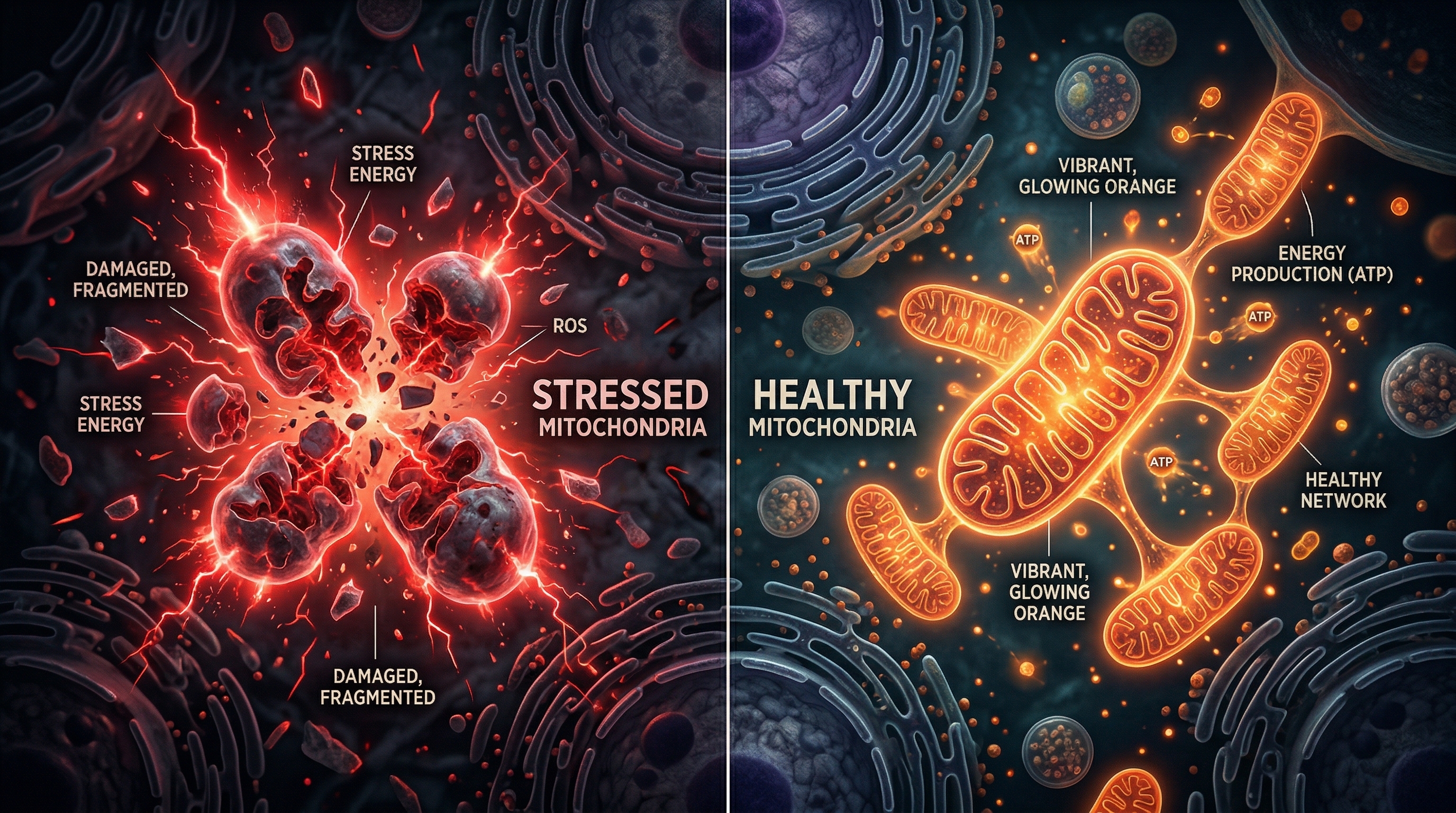
1. ROS Overproduction: The Electron Leak
Under normal conditions, mitochondria produce a small amount of reactive oxygen species (ROS) as a byproduct of electron transport — roughly 1–2% of total electron flow. This ROS serves as a signaling molecule at low levels. But chronic cortisol exposure disrupts the electron transport chain, particularly Complex I, causing electrons to "leak" from the chain before they reach their destination.
The result: superoxide radical production increases dramatically. These highly reactive molecules damage mitochondrial DNA (which is more vulnerable than nuclear DNA because it lacks histones and has limited repair mechanisms), oxidize mitochondrial proteins, and peroxidize the lipid membranes that maintain the organelle's structural integrity.
Research published in PubMed (PMID: 31216749) demonstrates that excess cortisol directly induces oxidative stress in hippocampal neurons, leading to structural mitochondrial damage visible on electron microscopy — swollen cristae, disrupted membranes, and fragmented organelles.
2. Mitochondrial Dynamics Disruption
Healthy mitochondria maintain a balance between fission (division) and fusion (merging). This isn't just housekeeping — it's how mitochondria share resources, distribute energy production, and isolate damaged components for repair or recycling (mitophagy).
Chronic stress skews this balance toward excessive fission. Research shows that glucocorticoid exposure upregulates Drp1 (dynamin-related protein 1) and Fis1 (fission protein 1) while simultaneously suppressing the fusion proteins OPA1, Mfn1, and Mfn2. The result: instead of a network of interconnected, efficient mitochondrial tubules, cells end up with fragmented, isolated mitochondria that are smaller, less efficient, and more prone to producing ROS.
3. Membrane Potential Collapse
The inner mitochondrial membrane maintains a voltage gradient of approximately -180 mV — a tiny but crucial electrical potential that drives ATP synthesis through ATP synthase. This membrane potential is maintained by the proton gradient generated during electron transport.
Oxidative stress from chronic cortisol exposure damages the lipid bilayer of the inner membrane, making it "leaky." Protons flow back across the membrane without passing through ATP synthase, dissipating the gradient. The mitochondrion must work harder to maintain the same ATP output, which generates more ROS, which causes more membrane damage — a self-amplifying cycle of decline.
When the damage becomes severe enough, the mitochondrial permeability transition pore (MPTP) opens — a catastrophic event that releases cytochrome c and activates apoptotic cascades. The cell dies. In neurons, cardiac muscle, and other post-mitotic tissues, this cell death is essentially irreversible.
4. Mitochondrial DNA Damage
Mitochondrial DNA (mtDNA) encodes 13 essential proteins of the electron transport chain, along with the tRNAs and rRNAs needed for their synthesis within the mitochondrion itself. Unlike nuclear DNA, mtDNA has no protective histone coating, limited mismatch repair, and sits in close proximity to the electron transport chain — right where ROS production is highest.
Chronic oxidative stress causes point mutations, deletions, and strand breaks in mtDNA. As these mutations accumulate, the affected mitochondria produce progressively less ATP and more ROS. Because each cell contains hundreds to thousands of mitochondria, this decline is gradual — until a threshold is crossed and cellular energy production falls below the level needed to sustain normal function.
5. Biogenesis Suppression
Under acute stress, cells respond by producing new mitochondria (mitochondrial biogenesis) to meet increased energy demands. This process is regulated by PGC-1α (peroxisome proliferator-activated receptor gamma coactivator 1-alpha), the master transcription factor for mitochondrial biogenesis.
Chronic stress suppresses PGC-1α expression. Research in a 2023 Frontiers in Physiology review confirmed that prolonged glucocorticoid exposure downregulates PGC-1α and its downstream targets (NRF1, NRF2, TFAM), reducing the cell's ability to replace damaged mitochondria. The result: a gradual decline in mitochondrial number and quality that manifests as fatigue, cognitive decline, and reduced physical capacity.
Stress → Cortisol → ROS ↑ → Mitochondrial damage → More ROS → Inflammatory signals → HPA activation → More cortisol. This feedback loop is why chronic stress accelerates biological aging and why simply "managing stress" is, in a very real sense, protecting your cellular power plants.
Who's Most at Risk?
While chronic stress affects everyone, certain populations face elevated mitochondrial risk:
- High-pressure professionals: Chronic work stress with sustained cortisol elevation
- Caregivers: Prolonged emotional stress combined with sleep disruption
- People with PTSD: A 2018 systematic review found that 19 out of 23 studies showed mitochondrial abnormalities in psychological stress conditions
- Those with depression: Emerging evidence links depressive episodes to measurable mitochondrial dysfunction
- Aging populations: Mitochondrial decline is a hallmark of aging; chronic stress accelerates it
What to Do About It: Practical Interventions
Tier 1: Reduce the Input
The most direct intervention is reducing chronic HPA activation:
- Vagus nerve activation: Slow diaphragmatic breathing (6 breaths per minute), cold exposure, and humming all stimulate the vagus nerve, which directly counteracts the stress response and activates the cholinergic anti-inflammatory pathway — protecting mitochondria from inflammatory damage.
- Sleep optimization: Cortisol follows a circadian rhythm. Poor sleep disrupts this rhythm, keeping cortisol elevated during periods when it should be low. Prioritizing 7–9 hours of quality sleep is arguably the single most impactful mitochondrial intervention available.
- Mindfulness and meditation: Regular meditation practice has been shown to improve heart rate variability (a marker of vagal tone) and reduce inflammatory cytokines. Research suggests it may also upregulate BDNF, which supports mitochondrial health in neurons.
Tier 2: Support the Mitochondria
Once you've reduced the stress input, directly support mitochondrial resilience:
- Exercise — but intelligently: Moderate aerobic exercise activates PGC-1α and stimulates mitochondrial biogenesis. But overtraining is itself a stressor that can damage mitochondria. Aim for consistent, moderate-intensity activity with adequate recovery.
- Nutritional support: CoQ10 (ubiquinol) directly supports electron transport. Magnesium is essential for ATP production. B vitamins serve as cofactors in mitochondrial metabolism. Omega-3 fatty acids reduce inflammatory ROS production.
- NAD+ precursors: Nicotinamide riboside (NR) and nicotinamide mononucleotide (NMN) support the NAD+ pool that mitochondria depend on for electron transport. Levels decline with age and chronic stress.
- Antioxidant support — targeted, not indiscriminate: N-acetylcysteine (NAC) supports glutathione production, your cells' primary endogenous antioxidant. But be cautious with high-dose exogenous antioxidants (vitamin C, vitamin E) — they may interfere with the beneficial ROS signaling that exercise and photobiomodulation rely on.
Tier 3: Repair the Damage
For those with established mitochondrial dysfunction:
- Red light therapy (photobiomodulation): Directly stimulates cytochrome c oxidase, enhancing electron transport and ATP production. Particularly effective for mitochondrial support because it generates mild, beneficial ROS that trigger protective adaptations without causing damage.
- Therapeutic ultrasound: Activates the vagus nerve and enhances cellular repair through mechanotransduction, supporting mitochondrial function through both neural and direct cellular pathways.
- Cold therapy: Beyond vagal activation, cold exposure stimulates mitochondrial biogenesis — your body creates more mitochondria to handle the metabolic demands of thermogenesis.
The Bottom Line
Stress isn't just a feeling — it's a biochemical event with physical consequences at the most fundamental level of your biology. Your mitochondria bear the brunt of chronic stress, and their decline drives many of the symptoms we associate with aging, burnout, and chronic disease.
The good news is that mitochondria are remarkably responsive to intervention. Reduce the stress signal, support their function, and they can recover. Your cells are always listening — the question is whether the signals they're receiving are telling them to survive or to surrender.
References
- PMC6627841. "Cortisol-induced oxidative stress and mitochondrial damage in hippocampal neurons."
- PubMed PMID: 31216749. "Excess cortisol and neuronal apoptosis via mitochondrial pathways."
- Psychoneuroendocrinology. "Glucocorticoid effects on mitochondrial oxidative phosphorylation." doi: 10.1016/j.psyneuen.2013.02.002
- PMC9220895. "Inverted U-shaped relationship between cortisol and mitochondrial function."
- International Journal of Molecular Sciences 25(7):3776. "Cortisol, ROS, and mitochondrial uncoupling in human platelets."
- PMC5901654. "Psychological stress and mitochondria: a systematic review."
- Biological Psychiatry: CNNI (2018). "Psychological stress and mitochondria." doi: 10.1093/bpych/bpy003
- Frontiers in Physiology (2023). "Acute stress, mitochondrial biogenesis, and hormesis." doi: 10.3389/fphys.2023.1222826
- PubMed PMID: 35757176. "NAC and mitochondrial protectors in stress-related dysfunction."