PEMF Therapy and Mitochondrial Function: Electromagnetic Fields at the Cellular Level
Pulsed Electromagnetic Field (PEMF) therapy has been FDA-approved for bone fracture healing since 1979, but the real story extends far deeper than bone repair. PEMF fields penetrate tissue and interact directly with cellular machinery — particularly mitochondria, the powerhouses of your cells.
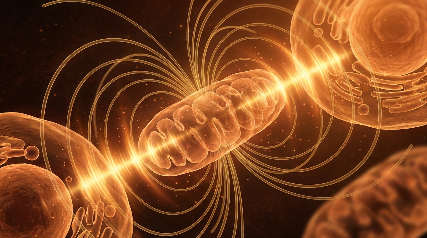
How PEMF Reaches Your Mitochondria
Unlike surface-level treatments, PEMF uses time-varying magnetic fields that pass through tissue without attenuation. These fields induce tiny electrical currents in cell membranes and — critically — in the mitochondrial membrane. The electromagnetic coupling is most effective at specific frequencies and intensities that match the natural resonance of biological structures.
Mitochondria are particularly susceptible to electromagnetic influence because of their:
- Double membrane structure: Creates capacitive coupling with oscillating fields
- Electron transport chain: An inherently electrical process that generates the proton gradient for ATP synthesis
- Calcium handling: Mitochondria are major calcium buffers, and calcium is highly sensitive to electromagnetic fields
ATP Production and the Electron Transport Chain
The electron transport chain (ETC) in the inner mitochondrial membrane is essentially a biological electrical circuit. Electrons pass through Complexes I, III, and IV, pumping protons across the membrane to create the gradient that drives ATP synthase. PEMF has been shown to enhance this process in several ways:
PEMF at 15-75 Hz and 1-5 mT has been shown to increase ATP production by 38-50% in multiple cell types, with peak effects occurring at 30-50 Hz.
The mechanism appears to involve improved electron flow through the ETC, reduced electron leakage (which generates damaging reactive oxygen species), and enhanced coupling efficiency of ATP synthase.
Calcium Signaling: The Second Messenger
Calcium ions (Ca²⁺) are the universal second messengers of cellular signaling. Mitochondria serve as calcium sinks, buffering cytoplasmic calcium levels and using calcium to regulate their own metabolic output. PEMF influences mitochondrial calcium handling through several pathways:
- Voltage-gated calcium channels: PEMF modulates the opening probability of these channels, altering calcium influx
- Mitochondrial calcium uniporter: PEMF enhances calcium uptake into the mitochondrial matrix
- Calcium-activated enzymes: Increased matrix calcium activates pyruvate dehydrogenase, isocitrate dehydrogenase, and α-ketoglutarate dehydrogenase — all rate-limiting enzymes in the TCA cycle
Clinical Evidence for PEMF and Energy
A growing body of clinical research supports PEMF's role in cellular energy optimization:
- Fatigue reduction: Multiple RCTs show PEMF reduces fatigue scores by 30-60% in chronic fatigue patients
- Exercise recovery: Athletes using PEMF post-training show faster muscle recovery and reduced oxidative stress markers
- Sleep quality: Low-frequency PEMF (1-10 Hz) improves sleep architecture and delta wave activity
- Pain management: PEMF reduces chronic pain through anti-inflammatory mechanisms linked to mitochondrial function
Practical Considerations
Not all PEMF devices are created equal. The therapeutic window for mitochondrial effects appears to be:
- Frequency: 1-75 Hz (cellular resonance range)
- Intensity: 0.5-5 mT (low intensity, non-thermal)
- Duration: 8-30 minutes per session
- Waveform: Bipolar square or sawtooth waves show highest efficacy
The future of PEMF lies in personalized protocols — matching frequency and intensity to individual mitochondrial phenotypes, creating a truly bioelectric approach to energy optimization.