Mitochondria and Brain Health: The Energy Behind Every Thought
Mitochondria and Brain Health: The Energy Behind Every Thought
Your brain weighs roughly three pounds — about 2% of your body mass. Yet it consumes approximately 20% of your body's total energy. This disproportionate metabolic demand means that your neurons are, gram for gram, the most energy-hungry cells you possess. And the mitochondria that supply that energy are not just supporting brain function — they are, in a very real sense, making thought itself possible.
The emerging field of neuroenergetics is revealing that mitochondrial dysfunction isn't just a consequence of brain disease. In many cases, it appears to be a cause — or at minimum, a critical early event that sets neurodegeneration in motion years or decades before symptoms appear.
The Neuron's Energy Problem
Neurons have a unique energy challenge. Unlike most cells, which can temporarily switch to anaerobic glycolysis when oxygen is limited, neurons depend almost entirely on oxidative phosphorylation. They cannot meaningfully store glucose or oxygen reserves. And their energy demands don't pause — the brain must maintain ion gradients across membranes even at rest, consuming ATP to run the Na⁺/K⁺-ATPase pumps that keep neurons electrically responsive.
Consider what a single neuron must power:
- Action potentials: Each time a neuron fires, it must rapidly restore sodium and potassium gradients across its membrane — an ATP-intensive process.
- Synaptic transmission: Neurotransmitter release, reuptake, and recycling all require energy. Synapses are the most metabolically expensive structures in the neuron.
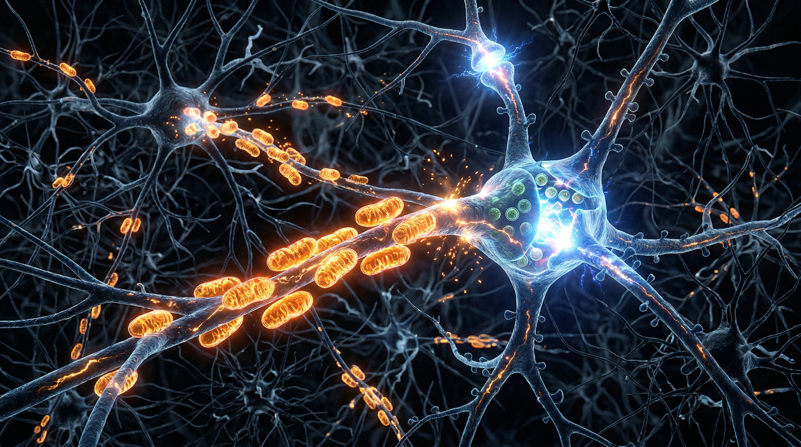
- Axonal transport: Mitochondria, proteins, and vesicles must be transported along axons that can extend over a meter in length (think: motor neurons running from your spine to your toes).
- Calcium homeostasis: Neurons use calcium as a signaling molecule and must carefully regulate cytoplasmic calcium levels through mitochondrial buffering and ER-mitochondria contact sites.
A 2022 review in FEBS Letters described mitochondrial dynamics — fission, fusion, transport, biogenesis, and mitophagy — as essential for maintaining energy supply along axons and dendrites. When these dynamics fail, the most distant synaptic terminals lose energy first, and it's at synapses that neurodegeneration often begins.
Mitochondrial Dynamics: The Brain's Quality Control System
Mitochondria in neurons are not static. They constantly undergo:
Fusion
Adjacent mitochondria merge their membranes, mixing contents and diluting damaged components. This process, mediated by Mitofusin 1/2 (MFN1/2) on the outer membrane and OPA1 on the inner membrane, allows functional mitochondria to compensate for partially damaged ones. Fusion is especially important in long axons, where mitochondria must travel far from the cell body.
Fission
Mitochondria divide through the action of Drp1 (dynamin-related protein 1). Fission allows the cell to segregate severely damaged mitochondria for recycling. But excessive fission — producing small, fragmented mitochondria — is a hallmark of neuronal stress and early neurodegeneration.
Transport
Neurons actively shuttle mitochondria along microtubule tracks to areas of high energy demand — particularly active synapses. This transport, driven by motor proteins kinesin and dynein, ensures that the places doing the most work get the most fuel. When transport fails, synapses starve.
Mitophagy
The PINK1/Parkin pathway tags damaged mitochondria for autophagic degradation. This quality control system is especially critical in post-mitotic cells like neurons, which cannot dilute damaged organelles through cell division. Impaired mitophagy leads to accumulation of dysfunctional mitochondria — and it's no coincidence that mutations in PINK1 and Parkin cause early-onset Parkinson's disease.
Alzheimer's Disease: An Energy Crisis Before a Protein Problem
One of the most compelling lines of evidence linking mitochondria to brain disease comes from Alzheimer's research. PET imaging studies have consistently shown that reduced glucose metabolism in the brain — called cerebral hypometabolism — can be detected years before cognitive symptoms appear, and it often precedes the formation of amyloid-beta plaques.
This temporal sequence matters. If energy failure comes before amyloid accumulation, it suggests that mitochondrial dysfunction may be an upstream driver of Alzheimer's pathology, not merely a downstream consequence.
Specific mitochondrial defects found in Alzheimer's brains include:
- Reduced activity of Complex IV (cytochrome c oxidase) in affected brain regions.
- Decreased expression of MFN2, impairing mitochondrial fusion and fragmenting the mitochondrial network.
- Amyloid-beta itself accumulating inside mitochondria, directly inhibiting ETC complexes and increasing ROS production.
- Impaired mitochondrial calcium buffering, leading to excitotoxic stress.
A bioenergetic hypothesis of Alzheimer's proposes that age-related decline in brain glucose uptake, NAD⁺/NADH ratio, and ETC efficiency creates a metabolic vulnerability — particularly in brain regions like the frontal cortex and temporoparietal cortex that have the highest baseline energy demands. When these regions can no longer sustain synaptic function, cognitive decline follows.
Parkinson's Disease: When Mitochondria Fail in the Substantia Nigra
Parkinson's disease offers perhaps the clearest genetic link between mitochondria and neurodegeneration. The dopaminergic neurons of the substantia nigra that degenerate in Parkinson's are uniquely vulnerable for several mitochondrial reasons:
- Complex I deficiency: Post-mortem studies consistently find reduced Complex I activity in the substantia nigra of Parkinson's patients. This is the same defect produced by MPTP, a neurotoxin that causes acute parkinsonism by specifically inhibiting Complex I.
- PINK1 and Parkin mutations: Autosomal recessive early-onset Parkinson's is caused by mutations in genes directly involved in mitochondrial quality control. Loss of PINK1 or Parkin function impairs mitophagy, allowing damaged mitochondria to accumulate in dopamine neurons.
- DJ-1 mutations: Another Parkinson's gene, DJ-1, protects mitochondria from oxidative stress. A recent Yale study confirmed that DJ-1 loss impairs ATP synthase function in distant neurons, explaining why Parkinson's pathology spreads along connected neural pathways.
- α-synuclein interaction: The protein aggregates characteristic of Parkinson's interact with mitochondria, impairing Complex I and disrupting mitochondrial membrane integrity.
ALS, Huntington's, and Beyond
The mitochondrial connection extends across the neurodegenerative spectrum. In ALS (amyotrophic lateral sclerosis), mutant SOD1 protein accumulates in mitochondria, disrupting ETC function and triggering apoptosis in motor neurons. In Huntington's disease, mutant huntingtin protein impairs mitochondrial dynamics and calcium handling, with striatal neurons — which have high metabolic demands — suffering preferentially.
Even in conditions not traditionally viewed as "neurodegenerative," mitochondrial health matters. Depression, anxiety, and cognitive aging are all associated with measurable changes in brain energy metabolism. The mitochondrion is emerging as a convergence point for understanding mental health at a biological level.
Protecting Brain Mitochondria: What the Evidence Shows
Exercise
Physical exercise upregulates brain-derived neurotrophic factor (BDNF), which promotes mitochondrial biogenesis in neurons through the PGC-1α pathway. Regular aerobic exercise increases hippocampal volume, improves synaptic plasticity, and reduces Alzheimer's risk — effects that are at least partly mediated by enhanced mitochondrial function.
Intermittent Fasting and Caloric Restriction
Fasting activates AMPK and SIRT1 in the brain, promoting mitochondrial quality control and biogenesis. Animal studies show that caloric restriction extends lifespan and reduces neurodegeneration — effects linked to improved mitochondrial function in cortical and hippocampal neurons.
NAD⁺ Precursors
Declining NAD⁺ levels are increasingly recognized as a central feature of brain aging. NMN and NR supplementation restores NAD⁺ in aged animal brains, improving mitochondrial function, synaptic plasticity, and cognitive performance. Human clinical trials are ongoing, with early results showing promise for safety and bioavailability.
Antioxidant Support
While general antioxidant supplementation has shown mixed results in neurodegeneration trials, mitochondria-targeted antioxidants like MitoQ (which accumulates specifically in mitochondria) have shown benefits in preclinical models of Alzheimer's and Parkinson's disease.
The essential insight: Every thought you think, every memory you form, every emotion you feel is powered by mitochondrial ATP. When brain mitochondria fail — whether through aging, genetic vulnerability, or environmental stress — the consequences reach far beyond fatigue. They reshape cognition, mood, and the trajectory of your entire neurological life. Protecting your mitochondria isn't just a longevity strategy. It's a brain health strategy.
References
- Mishra P, Chan DC. "Metabolic Regulation of Mitochondrial Dynamics." Journal of Cell Biology, 2016;212(4):379-387.
- Swerdlow RH. "The Neuroenergetic Theory of Brain Aging." Frontiers in Aging Neuroscience, 2023;15:1153414.
- Picard M, McEwen BS. "Psychological Stress and Mitochondria: A Systematic Review." Psychosomatic Medicine, 2018;80(2):141-153.
- Devine MJ, Kittler JT. "Mitochondria at the Neuronal Presynapse in Health and Disease." Nature Reviews Neuroscience, 2018;19(2):63-80.
- Golpich M et al. "Mitochondrial Dysfunction and Biogenesis in Neurodegenerative Diseases." ACS Chemical Neuroscience, 2017;8(6):1185-1203.
- Yao J et al. "Mitochondrial Targeted Therapy for Alzheimer's Disease." Journal of Alzheimer's Disease, 2020;76(1):1-20.
- Valenti D et al. "Mitochondrial Dysfunction in Parkinson's Disease." FEBS Letters, 2022;596:1476-1493.
- Yin F et al. "Energy Metabolism and Inflammation in Brain Aging and Alzheimer's Disease." Free Radical Biology and Medicine, 2016;100:108-122.