Tinnitus and the Vagus Nerve: Why the Phantom Sound Lives in Your Gain, Not Your Ear
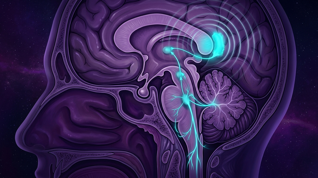
Most people with chronic tinnitus are told the same thing: the damage is in the ear, there is no cure, learn to live with it. But the ear is rarely where the sound is generated. When I look closely at the phantom-sound patients who never respond to hearing aids or masking alone, the pattern is consistent — their tinnitus is loud when they are stressed, wired at night, and running low on sleep, and quieter on the rare calm morning. That is not the signature of a fixed cochlear lesion. It is the signature of a nervous system that has turned up its own gain. Tinnitus, in the majority of persistent cases, is a central gating problem, and the vagus nerve sits close to the controls.
The Ear Loses Input, the Brain Turns Up the Volume
Almost every case of chronic tinnitus begins with some reduction in cochlear input — noise exposure, age-related high-frequency loss, an ototoxic drug, a viral insult, sometimes a loss too subtle to show on a standard audiogram. The intuitive expectation is that less input should mean less sound. The opposite happens. When a frequency channel goes quiet, the central auditory pathway does not sit idle; it compensates.
This is central gain. Deprived of their normal afferent drive, neurons in the dorsal cochlear nucleus, inferior colliculus, and auditory thalamus increase their spontaneous firing and their responsiveness. The system is behaving like an audio engineer who cranks the amplifier to hear a fading signal — and in doing so, amplifies the noise floor into an audible tone. The phantom sound is the amplified hiss of a pathway compensating for lost input. Homeostatic mechanisms that normally keep neural excitability in a healthy band overshoot, inhibition (particularly GABAergic tone) weakens, and the gain never resets.
Thalamocortical Dysrhythmia: The Phantom Gets a Rhythm
Gain alone produces noise. What turns noise into a stable, perceived tone is a rhythm problem one level up. In a healthy auditory thalamocortical loop, thalamic relay cells fire in a fast, desynchronized mode that faithfully passes sound to the cortex. When those cells are deprived of input, they hyperpolarize and switch into slow burst-firing driven by T-type calcium channels — a low-frequency theta rhythm.
That slow thalamic rhythm entrains a patch of auditory cortex. Around the edge of the abnormally slow region, the normal lateral inhibition breaks down and a ring of high-frequency gamma activity appears — and gamma in auditory cortex is heard as sound. This is the thalamocortical dysrhythmia model, and it explains why tinnitus is so often a steady tone rather than random static: the phantom has acquired a pathological rhythm. It also explains why tinnitus is genuinely perceived. The person is not imagining it; their auditory cortex is producing the neural correlate of a sound that has no acoustic source.
Who Sets the Gain? The Locus Coeruleus, Acetylcholine, and the Vagus
Here is the part conventional otology tends to skip, and the part that matters most for a coach. Central gain is not a fixed dial set by the cochlea. It is state-dependent, set moment to moment by ascending neuromodulatory systems — and those systems are exactly the ones the autonomic nervous system and the vagus nerve govern.
Two neuromodulators dominate auditory gain. Noradrenaline, released from the locus coeruleus, is the brain's arousal and threat-salience signal; it sharpens sensory responsiveness and biases the system toward vigilance. Acetylcholine, from the basal forebrain, gates cortical plasticity and attention. When the locus coeruleus is chronically overactive — the neurochemical reality of chronic stress, hypervigilance, and poor sleep — auditory neurons are held in a high-gain, high-alert state. The amplifier is turned up by arousal, independent of anything happening in the ear.
The vagus nerve is the counterweight. Its afferent fibers — roughly eighty percent of the nerve — carry interoceptive information from the heart, lungs, and gut to the nucleus tractus solitarius (NTS) in the brainstem. From the NTS, projections reach the locus coeruleus, the raphe nuclei, the parabrachial nucleus, and onward to the amygdala and insula. This is why the vagus is not a bystander to auditory gain: vagal afferent tone directly modulates the locus coeruleus and the cholinergic and serotonergic systems that set sensory excitability. High vagal tone biases the brainstem toward a low-arousal, low-gain, parasympathetic-dominant state. Low vagal tone releases the brake, the locus coeruleus runs hot, noradrenergic gain climbs, and the phantom gets louder. This is the mechanistic bridge between a patient's heart-rate variability and the volume of a sound only they can hear.
The Limbic-Autonomic Amplification Loop
Gain explains the sound. It does not explain the suffering, and the two dissociate completely — some people have measurable tinnitus and barely notice it, while others are disabled by a sound of identical pitch and intensity. The difference is a second loop layered on the first.
When the auditory phantom is flagged as threatening, the amygdala and anterior insula attach emotional and interoceptive salience to it. The amygdala projects back down to the brainstem and drives sympathetic activation and further locus coeruleus firing — which raises auditory gain — which makes the sound more salient — which feeds the amygdala. This is a self-reinforcing limbic-autonomic loop: threat raises gain, gain raises salience, salience confirms threat. The tinnitus becomes both louder and more distressing over time not because the ear is deteriorating, but because the loop is consolidating. Sleep loss pours fuel on it, because sleep deprivation alone elevates locus coeruleus tone and lowers vagal tone. Patients accurately report that a bad night makes the sound roar the next day.
The Autonomic Phenotype: Recognizing Central Tinnitus
This mechanism produces a recognizable clinical picture that a coach can learn to spot:
- Tinnitus loudness that tracks stress, sleep, and arousal more tightly than any acoustic exposure.
- Hyperacusis — ordinary sounds perceived as too loud or painful — riding alongside the tinnitus. Both are the same central gain expressed on external versus internal input.
- Broader sensory hypersensitivity: to light, touch, or busy visual environments, pointing to a whole nervous system biased toward threat.
- Comorbid anxiety, insomnia, and a low resting heart-rate variability.
- Onset clustered around a stressor or a viral illness rather than a single acoustic trauma. Post-viral tinnitus, including after COVID, fits this pattern: viral and inflammatory insults hit both the auditory periphery and autonomic control, and the dysautonomia that follows keeps the gain elevated.
When these features cluster, the useful reframe is not "your ears are damaged" but "your nervous system is stuck in a high-gain, high-threat state, and the sound is what that state sounds like." That reframe is not a consolation. It names two things — vagal tone and autonomic arousal — that are genuinely modifiable.
When to Get Evaluated: Red Flags
The central-gain model applies to the common bilateral, non-pulsatile, chronic tinnitus that dominates clinic populations. It does not apply to everything, and some presentations need urgent medical evaluation before any nervous-system work. See a physician or ENT promptly if the tinnitus is:
- Unilateral (one ear only) or clearly asymmetric — this can signal a retrocochlear lesion such as a vestibular schwannoma and warrants imaging.
- Pulsatile — beating in time with the heartbeat — which can reflect a vascular abnormality and needs a specific workup.
- Accompanied by sudden hearing loss, which is a medical emergency; prompt treatment materially changes outcomes.
- Paired with vertigo, neurological signs, or a recent head injury.
Clearing these first is not a formality. It is what makes the reassurance underlying every nervous-system intervention honest.
Where Neuromodulation and Ultrasound Fit
Tinnitus is one of the few areas where vagal neuromodulation has moved from theory into active clinical research, precisely because the vagus modulates the neuromodulators that set auditory gain and plasticity.
The most developed approach is paired vagus nerve stimulation. The logic is elegant: acetylcholine and noradrenaline released by vagal stimulation open a window of cortical plasticity, so pairing a brief VNS pulse with a tone other than the tinnitus frequency can, over many repetitions, nudge the over-represented cortical map back toward normal and shrink the phantom. Early implanted-VNS work established the principle, and the field has since moved decisively toward non-invasive bimodal stimulation — pairing sound with mild electrical stimulation of a peripheral nerve. Large studies of a bimodal device delivering tones alongside tongue stimulation (the Lenire line of research) have reported meaningful reductions in tinnitus severity, and the through-line is the same: drive the paired neuromodulatory release that reorganizes the maladaptive map.
Transcutaneous auricular VNS (taVNS) is the accessible cousin. The outer ear's concha is one of the few places a branch of the vagus reaches the skin, so a small electrode there stimulates vagal afferents directly into the NTS and up to the locus coeruleus — turning down noradrenergic gain without surgery. taVNS for tinnitus is early-stage and results are mixed, but the mechanistic rationale is sound and the research is genuinely ongoing.
Low-intensity focused ultrasound is the emerging frontier. Because ultrasound can be focused non-invasively onto deep targets, it is being studied as a way to neuromodulate the cervical vagus and, potentially, the thalamic and limbic nodes that generate dysrhythmia — reaching structures that surface electrodes cannot. This work is early; we expect the clearest evidence to arrive over the coming years, and the honest framing today is "promising and being actively studied," not "proven."
None of this requires a device to begin. Every mechanism above is also reachable through the vagal training a coach can teach directly. Slow breathing at roughly six breaths per minute with long exhales raises vagal afferent traffic and lowers locus coeruleus arousal — the same gain dial the implanted devices target. Humming, gargling, and singing mechanically engage the vagally-innervated laryngeal and pharyngeal muscles and are, conveniently, also masking sound. Cold-water face immersion triggers the dive reflex, a fast and powerful parasympathetic surge. HRV biofeedback gives the patient a live readout of vagal tone and a way to learn the low-gain state on demand. And protecting sleep is not adjunctive — it is a primary intervention, because sleep is when locus coeruleus tone falls and the amplifier resets.
Clinical takeaway: Treat chronic non-pulsatile tinnitus as a central-gain and autonomic-gating problem first, an ear problem second. The cochlea may have set the stage, but the loudness and the distress are governed by locus coeruleus arousal and vagal tone — both trainable. Coach the vagus (slow exhales, humming, cold, HRV biofeedback, sleep) to lower the amplifier, and understand paired-VNS and taVNS as the same lever applied with hardware. Rule out unilateral, pulsatile, or sudden-onset presentations first.
References
- Eggermont JJ, Roberts LE. "The neuroscience of tinnitus." Trends in Neurosciences, 2004;27(11):676-682.
- Roberts LE, Eggermont JJ, Caspary DM, et al. "Ringing ears: the neuroscience of tinnitus." Journal of Neuroscience, 2010;30(45):14972-14979.
- Llinás RR, Ribary U, Jeanmonod D, et al. "Thalamocortical dysrhythmia: A neurological and neuropsychiatric syndrome characterized by magnetoencephalography." Proceedings of the National Academy of Sciences, 1999;96(26):15222-15227.
- De Ridder D, Vanneste S, Weisz N, et al. "An integrative model of auditory phantom perception: Tinnitus as a unified percept of interacting separable subnetworks." Neuroscience & Biobehavioral Reviews, 2014;44:16-32.
- Engineer ND, Riley JR, Seale JD, et al. "Reversing pathological neural activity using targeted plasticity." Nature, 2011;470(7332):101-104.
- Conlon B, Langguth B, Hamilton C, et al. "Bimodal neuromodulation combining sound and tongue stimulation reduces tinnitus symptoms in a large randomized clinical study." Science Translational Medicine, 2020;12(564):eabb2830.
- Berthoud HR, Neuhuber WL. "Functional and chemical anatomy of the afferent vagal system." Autonomic Neuroscience, 2000;85(1-3):1-17.